Overview
Contact
What Is Medial Patellofemoral Ligament Reconstruction?

The patellofemoral joint is formed by the patella (kneecap) articulating within the trochlear groove of the distal femur, facilitating knee extension and load distribution during activities like squatting, running, and stair climbing. The medial patellofemoral ligament (MPFL) is a key static stabilizer, originating from the medial femoral condyle (between the adductor tubercle and medial epicondyle) and inserting onto the superomedial border of the patella. It provides 50-60% of the medial restraint against lateral patellar translation, particularly in the first 0-30 degrees of knee flexion when the trochlear groove offers less bony containment. Complementary structures include the vastus medialis obliquus (VMO) muscle for dynamic medial pull, the medial patellotibial ligament, and the trochlear dysplasia or patella alta that can influence overall stability. The quadriceps tendon superiorly and patellar tendon inferiorly further guide patellar tracking.
MPFL disruption commonly occurs during acute lateral patellar dislocations, often from non-contact twisting mechanisms in sports (e.g., soccer, basketball) or trauma, where the ligament tears at its femoral attachment (60-80% of cases) or mid-substance. This leads to altered patellar tracking, recurrent instability, and symptoms like apprehension, giving way, effusion, and pain during pivoting or extension. Associated factors include trochlear dysplasia (shallow groove), patella alta (high-riding patella), increased Q-angle, or ligamentous laxity, exacerbating lateral forces. Untreated, recurrent dislocations (up to 50% after first episode) cause progressive chondral damage to the patella or trochlea, medial patellofemoral osteoarthritis, and chronic pain or functional limitations. Clinical signs include a positive patellar apprehension test, J-sign (lateral patellar deviation in extension), and MRI-confirmed MPFL tear or bone bruises.
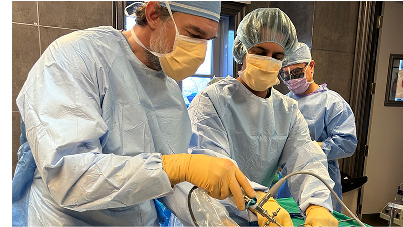
At OV Surgical, we elevate MPFL reconstruction in Canada with advanced, anatomic techniques that minimize downtime and maximize results. Performed under general or regional anesthesia with fluoroscopic guidance for precision, the procedure begins with diagnostic arthroscopy through standard portals to address intra-articular pathology like loose bodies or cartilage defects. A 2-3 cm incision over the medial patella exposes the graft harvest site—typically semitendinosus or gracilis autograft (or allograft for select cases)—which is prepared into a double-looped construct. Small incisions at the femoral origin (Schöttle point: 1 mm proximal to the adductor tubercle, 2.5 mm distal to the posterior cortex) and patellar insertion allow tunnel drilling: two small all suture anchors into the medial patella (avoiding articular violation) and a socket or full tunnel at the femur. The graft is passed subcutaneously, fixed patella-side anchors, and femur-side with a button or screw, tensioned at 30 degrees of flexion to restore isometry without overconstraint (2-3 mm lateral glide allowed). In cases of malalignment, adjunct tibial tubercle osteotomy medializes the tubercle. The surgery typically lasts 60-90 minutes, with meticulous closure to prevent hematoma and emphasis on preserving the VMO and neurovascular structures like the saphenous nerve.
Recovery
Post-procedure, our evidence-based protocols integrate cryotherapy, neuromuscular electrical stimulation (NMES), and progressive physiotherapy to accelerate healing and restore function, tailored to alignment corrections. In the immediate protection phase (0-6 weeks), patients use a hinged brace locked in extension for ambulation, with partial weight-bearing on crutches for 4-6 weeks. Passive range of motion (PROM) starts day 1 via continuous passive motion (CPM) machine or physiotherapist, aiming for 0-90 degrees flexion by week 4, alongside isometric quadriceps activations, straight-leg raises, and patellar mobilizations to prevent adhesions. Pain management includes ice and multimodal analgesia to control swelling.
From weeks 6-12 (active strengthening phase), you will progress to full ROM, and closed-chain exercises like partial squats or leg presses (2-3 sets of 10-15 reps), focusing on VMO recruitment to enhance dynamic stability. Gait normalization and balance drills on stable surfaces build proprioception while avoiding pivoting. By weeks 12-16 (functional strengthening phase), full active ROM is targeted with resistance training using bands or weights for quadriceps, hamstrings, and hip abductors, incorporating single-leg balances and step-ups to mimic daily activities. Advanced phases (16+ weeks) include agility ladders, plyometrics (e.g., box jumps), and sport-specific drills (e.g., cutting for athletes), emphasizing eccentric control. Most patients achieve full motion by 8-12 weeks and strength milestones by 3-4 months, with sports clearance in 6 months, verified through rigorous testing like quadriceps strength assessments (>90% symmetry), single-leg hops, patellar stability exams, and patient-reported outcomes (e.g., Kujala score). We emphasize psychological readiness, addressing fear of re-dislocation through gradual exposure and confidence-building.

Benefits
With success rates of 85-95%, private MPFL reconstruction at OV Surgical restores patellar stability, prevents re-dislocation, and allows you to get back to sports and/or other aspects of life that are important to you. Our patients report enhanced knee function, confidence, and quality of life, backed by meticulous follow-up and data-driven protocols.
FAQ
Contact