Overview
Contact
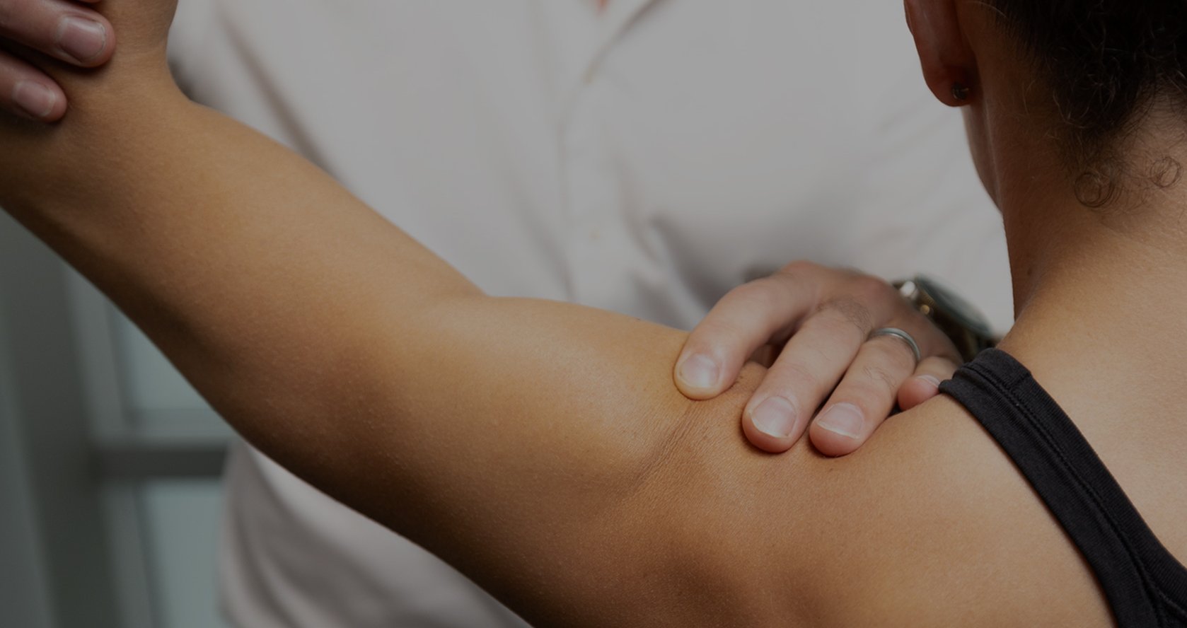
What Is Subacromial Decompression Surgery?
The shoulder is a highly mobile ball-and-socket joint formed by the humeral head articulating with the glenoid cavity of the scapula, supported by the clavicle. Key structures include the rotator cuff—a group of four tendons (supraspinatus, infraspinatus, teres minor, subscapularis) that stabilize the humeral head and enable elevation and rotation—and the subacromial space, a narrow gap between the acromion (the bony projection of the scapula) and the rotator cuff. Within this space lies the subacromial bursa, a fluid-filled sac that reduces friction during arm movement, and the coracoacromial (CA) ligament, which forms an arch over the rotator cuff.
Pathoanatomy of subacromial impingement syndrome involves narrowing of the subacromial space, leading to mechanical compression of the rotator cuff tendons and bursa against the acromion or CA ligament. This often results from bone spurs (osteophytes) on the undersurface of the acromion, thickening of the CA ligament, or age-related degenerative changes that reduce tendon vascularity and collagen integrity. Causes include repetitive overhead activities (e.g., in painters, swimmers, or tennis players), acute trauma, or intrinsic factors like rotator cuff tendinopathy, bursitis, or partial tears. Symptoms manifest as shoulder pain aggravated by overhead reaching, a painful arc between 60° and 120° of abduction, nocturnal (night) discomfort, weakness, and positive impingement tests (e.g., relief with subacromial lidocaine injection). Untreated, it can progress to chronic inflammation, fibrosis, full-thickness rotator cuff tears, or glenohumeral osteoarthritis.
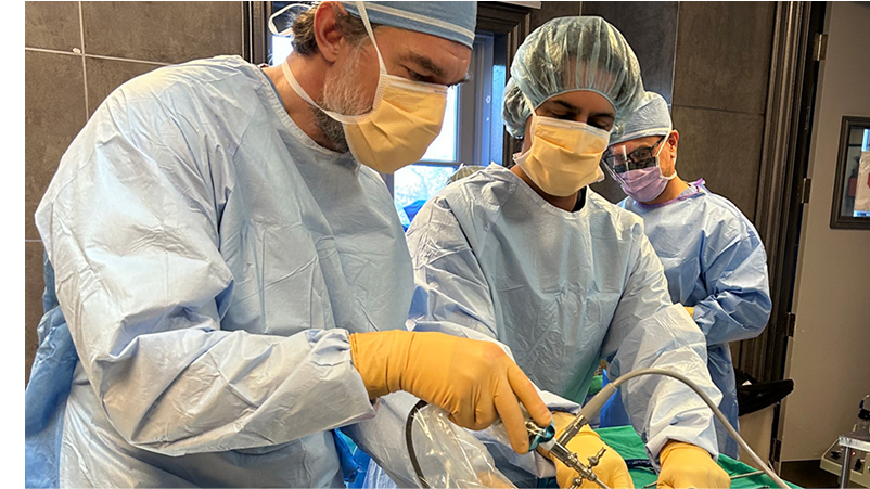
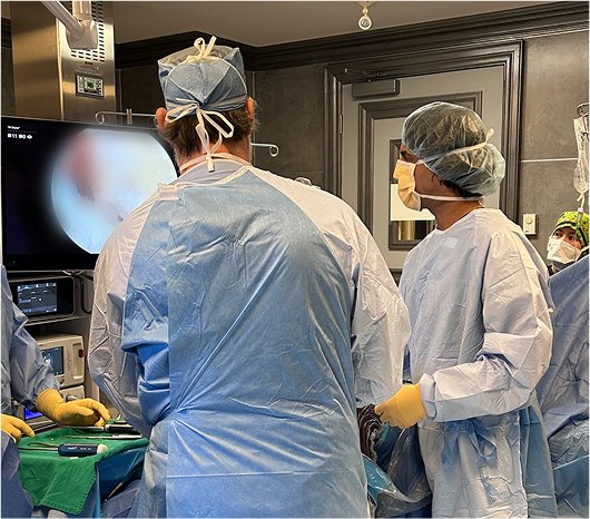
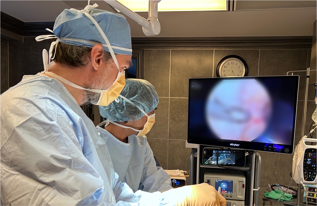
At OV Surgical, we advance subacromial decompression surgery in Canada with arthroscopic techniques that minimize downtime and optimize results. Performed under general anesthesia with possible interscalene block, the procedure uses two to three small incisions (less than 1 cm) to insert an arthroscope for visualization of the glenohumeral joint and subacromial space. Fluid inflates the joint for better access. Steps include bursectomy—removal of the inflamed subacromial bursa using a shaver or electrocautery to eliminate edematous tissue—and acromioplasty, where a motorized burr shaves 4-6 mm of bone from the anterior and lateral undersurface of the acromion to flatten it and widen the space. The CA ligament may be released if it contributes to impingement. Associated issues, like partial rotator cuff debridement or distal clavicle resection, are addressed if present. The surgery lasts 30-60 minutes, with incisions closed using absorbable sutures or steri-strips.
Recovery
Our evidence-based protocols use cryotherapy, neuromuscular electrical stimulation (NMES), and progressive physiotherapy, tailored to individual pain levels and tissue healing. In the protection phase (0-2 weeks), patients wear a sling for comfort (typically 1-4 days, up to 2 weeks if combined procedures), focusing on pain control with ice, anti-inflammatories, and prescribed medications. Passive range of motion (PROM) begins immediately to prevent stiffness, including pendulum exercises, standing scapular mobility, passive external rotation, flexion, internal rotation, and horizontal adduction (1-2 sets of 10-20 reps, 3-5 times daily). Wounds are kept dry for 5-7 days.
From weeks 2-6 (active ROM phase), the sling is discontinued, and active-assisted range of motion (AAROM) progresses with supine stick flexion, table/wall slides, and gentle cross-body stretches. Strengthening starts against gravity with prone rows, extensions, horizontal abductions, and sidelying external rotations (2-3 sets of 15-20 reps daily). Precautions include avoiding overhead lifting or repetitive use. By weeks 6-12 (strengthening phase), full active ROM is targeted with resistance bands or dumbbells for rows, scaption, internal/external rotations, and manual rhythmic stabilizations (3-5 times weekly, 2-3 sets of 15-20 reps). Proprioceptive drills at 90° elevation enhance control, with no lifting over 1-2 pounds overhead. Advanced phases (12+ weeks) incorporate diagonal patterns, plyometrics, push-up progressions, and sport-specific drills (e.g., throwing or overhead simulations), with gym lifting cleared by the surgeon. Full motion is typically achieved by 4-6 weeks, strength milestones by 8-12 weeks, and return to overhead work or sports by 3-6 months, verified through functional tests like impingement signs and strength symmetry (>90%). Psychological readiness addresses activity-related fear through gradual progression.
Benefits
With success rates of 80-90% in reducing pain and improving function (e.g., 80% symptom improvement by 3 months, full recovery within 4-6 months), private subacromial decompression at OV Surgical relieves impingement, prevents progression to rotator cuff tears, and supports return to sports and daily life. Patients report enhanced function, confidence, and quality of life, backed by meticulous follow-up and data-driven protocols.
FAQ
Contact